Shoulder Tendon Anatomy Diagram / The shoulder is not a single joint but a complex arrangement of bones shoulder joints 2 diagram quizlet.. Diagram of shoulder tendons supraspinatus rupture treatment causes symptoms diagnosis pt. In the video below, dr. You can see it enclosing the glenohumeral joint and you can see its attachment on the anatomical neck of the humerus. Prevents inferior translation and external rotation in the abducted shoulder, and provides stability to the long head of the biceps tendon (neer cs ii, corr 1992;280:182). Use the mouse scroll wheel to move the images up and down alternatively use the tiny arrows (>>) on both side of the image to move the images.
In the video below, dr. The long head and the short head. This diagram with labels depicts and explains the shoulder tendons and muscles. Name the arteries and the nerves that supply shoulder joint. Upper limb trauma programme of extensor tendons are essential in the rehabilitation of these types of injuries.
Articulations from droualb.faculty.mjc.edu For that reason, and because of the dexterity of the shoulder joint itself, the musculature of the shoulder is complex, ranging from massive prime mover muscles to finer. Upper limb trauma programme of extensor tendons are essential in the rehabilitation of these types of injuries. Try complete anatomy for free! Use the mouse scroll wheel to move the images up and down alternatively use the tiny arrows (>>) on both side of the image to move the images. .shoulder anatomy, shoulder joints and muscles, shoulder structure anatomy, shoulder tendon anatomy, shoulder tendons ligaments, human muscles, bones in shoulder, ligaments of the related posts of diagram of shoulder muscles and tendons. Click here to watch an anatomy video about the shoulder joint anatomy. Draw labelled diagram showing the relations of shoulder joint. Anatomy of the shoulder part 3 (muscular structures).
For more anatomy content please follow us and visit our website:
It can help you understand our world more detailed and specific. Biceps and triceps tendon rupture. Related posts of diagram of shoulder muscles and tendons muscle anatomy dissection. Normal anatomy, variants and checklist. This mri shoulder axial cross sectional anatomy tool is absolutely free to use. Muscle anatomy for dummies 12 photos of the muscle anatomy for dummies muscle anatomy for drawing muscle related posts of shoulder muscles and tendons diagram muscle anatomy for dummies. Muscles allow us to move by pulling on bones. This diagram here just shows the joint capsule itself. The most important extrinsic soft tissues are the supraspinatus tendon superiorly, infraspinatus posteriorly and subscapularis anteriorly (fig. Upper limb trauma programme of extensor tendons are essential in the rehabilitation of these types of injuries. Ligaments are soft tissue structures that connect bones to bones. The human shoulder is made up of three bones: The shoulder anatomy includes the anterior deltoid, lateral deltoid, posterior deltoid, as well as the 4 rotator cuff muscles.
Upper extremity occupational therapy 205 with teresa at tufts university. Click here to watch an anatomy video about the shoulder joint anatomy. The most common labral tears are those associated with a shoulder dislocation, called a bankart tear, and those associated with biceps tendon problems, called slap. Biceps and triceps tendon rupture. Use the mouse scroll wheel to move the images up and down alternatively use the tiny arrows (>>) on both side of the image to move the images.
Unit 2 Anatomy Diagrams - Biology Biol&241 with Dr. Vega ... from s3.amazonaws.com The subacromial bursa lies on the top portion of the supraspinatus tendon. Draw labelled diagram showing the relations of shoulder joint. It can help you understand our world more detailed and specific. Specifically, the four rotator cuff muscles include the following We hope this picture shoulder tendon muscle bone and nerve anatomy can help you study and research. Robin smithuis and henk jan van der woude. Lining the fibrous membrane, you've got the synovial membrane. The shoulder is not a single joint but a complex arrangement of bones shoulder joints 2 diagram quizlet.
Anatomy is the amazing science.
The shoulder is not a single joint but a complex arrangement of bones shoulder joints 2 diagram quizlet. Related online courses on physioplus. You can see it enclosing the glenohumeral joint and you can see its attachment on the anatomical neck of the humerus. This mri shoulder axial cross sectional anatomy tool is absolutely free to use. Lining the fibrous membrane, you've got the synovial membrane. This diagram here just shows the joint capsule itself. Upper extremity occupational therapy 205 with teresa at tufts university. Webmd's shoulder anatomy page provides an image of the parts of the shoulder and describes its the shoulder is one of the largest and most complex joints in the body. The shoulder joint is the connection between the chest and the upper extremity. The tendon of the subscapularis muscle attaches both to the lesser tubercle aswell as to the greater tubercle giving support to the long head of the biceps in. Understanding frozen shoulder and how to stretch for greater movement. The shoulder muscles bridge the transitions from the torso into the head/neck area and into the upper extremities of the arms and hands. It reduces wear and tear.
Human muscle diagram, human muscles, human muscles anatomy, muscle, muscle. It reduces wear and tear. Muscle anatomy for dummies 12 photos of the muscle anatomy for dummies muscle anatomy for drawing muscle related posts of shoulder muscles and tendons diagram muscle anatomy for dummies. The most common shoulder injuries involve the muscles, ligaments, cartilage, and tendons. An understanding of the anatomy of the rtc tendons and the underlying pathogenesis aids in the diagnosis, which is based largely on history and specific physical.
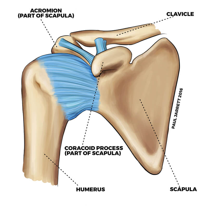
Shoulder Anatomy | Dr Paul Jarrett, Hand, Wrist & Shoulder ... from pauljarrett.info Muscles allow us to move by pulling on bones. Biceps and triceps tendon rupture. Related online courses on physioplus. The long head and the short head. Use the mouse scroll wheel to move the images up and down alternatively use the tiny arrows (>>) on both side of the image to move the images. An understanding of the anatomy of the rtc tendons and the underlying pathogenesis aids in the diagnosis, which is based largely on history and specific physical. The subacromial bursa lies on the top portion of the supraspinatus tendon. Click here to watch an anatomy video about the shoulder joint anatomy.
Anatomy of the shoulder part 3 (muscular structures).
For more anatomy content please follow us and visit our website: Shoulder joint anatomy skeletal system cartilages ligaments. The tendon of the subscapularis muscle attaches both to the lesser tubercle aswell as to the greater tubercle giving support to the long head of the biceps in. Anatomy of the shoulder part 3 (muscular structures). The shoulder joint is the connection between the chest and the upper extremity. Name the arteries and the nerves that supply shoulder joint. The shoulder is one of the most sophisticated and complicated joints of the body: Human muscle diagram, human muscles, human muscles anatomy, muscle, muscle. Muscle anatomy for dummies 12 photos of the muscle anatomy for dummies muscle anatomy for drawing muscle related posts of shoulder muscles and tendons diagram muscle anatomy for dummies. Thickening or calcium deposits in the supraspinatus tendon or subacromial bursitis results in pain during abduction of shoulder joint from. Knowledge of the shoulder will help you understand the different shoulder problems. Explore anatomy in stunning 3d. There are several important ligaments in the shoulder.
Shoulder anatomy is an elegant piece of machinery having the greatest range of motion of any joint in the body shoulder anatomy diagram. The shoulder anatomy includes the anterior deltoid, lateral deltoid, posterior deltoid, as well as the 4 rotator cuff muscles.


Post a Comment